

Research performed in veterinary medicine impacts companion and agricultural animals, as well as human health through the use of pre-clinical models of disease. Pursuits in this field range from understanding the impact of pollutants on various aspects of physiology to the response of the body to infectious agents of disease.
Recent advancements in light microscopy techniques and other new technologies have permitted the ability to observe specific structures that are smaller than a cell in an entire intact organ. This ability to observe cells in such unprecedented detail creates great opportunity to advance research with animals – but it also requires labor intensive processing of large data sets that slows the ability to make novel discoveries.
A data science approach that can deal with these types of data on this scale, as well as the development of new data analysis methods, will make the process of extracting quantifiable data from these large data sets quicker and more streamlined – increasing the rate of novel discoveries.
Regulation of neuronal development and behavior
Colin Reardon
The limbic system is important for modulating emotions and memories and continues to develop well after birth. Hippocampal volume, for example, continues to grow rapidly until age 2 in humans, after which growth decreases substantially. This process is coupled to the formation of new neurons, termed neurogenesis, occurs throughout life in specific brain regions including the hippocampus. The level of neurogenesis and memory persistence are causally related, with elevated neurogenesis in neonates associated with infantile amnesia.
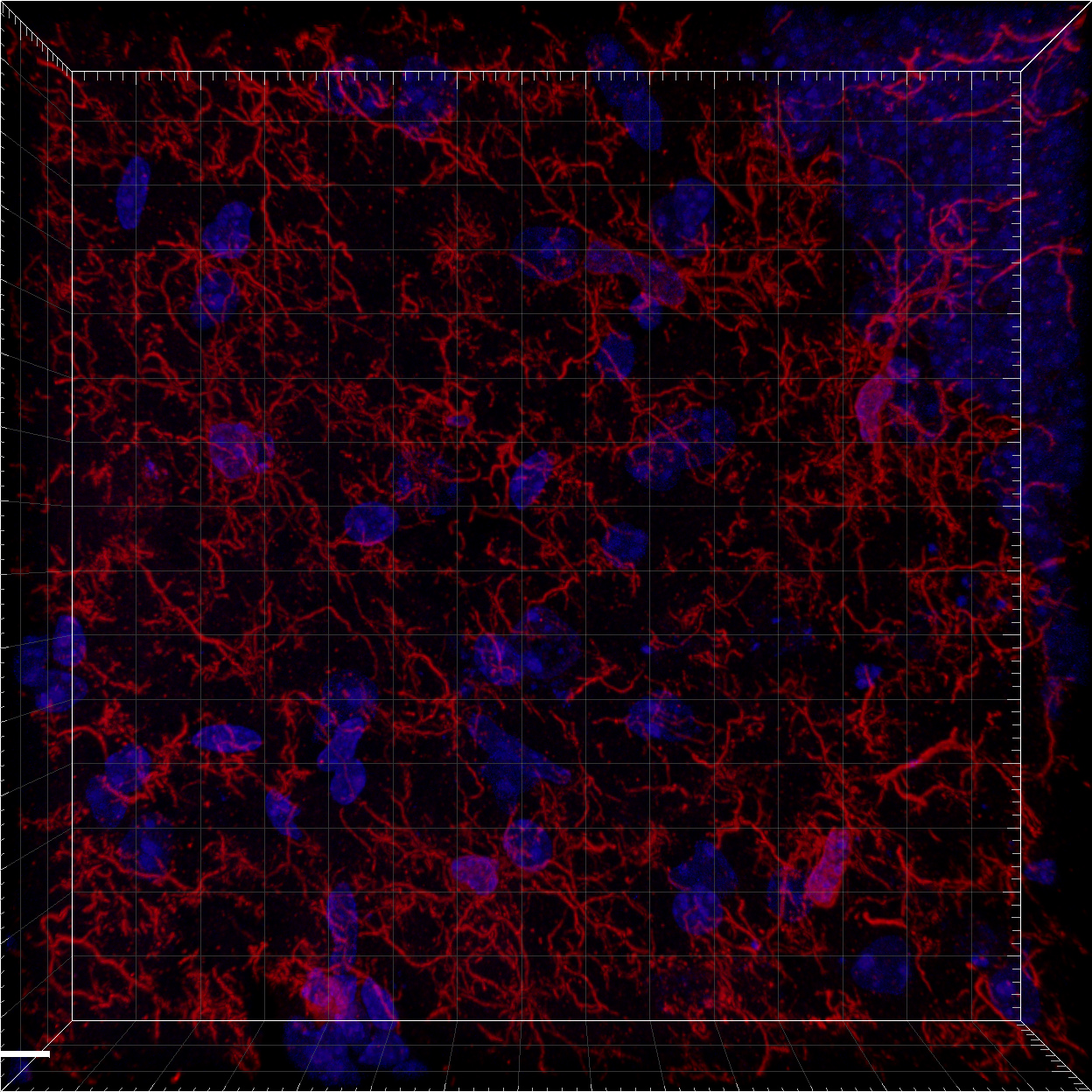
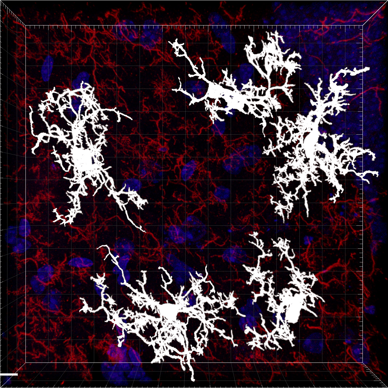
It is known that specialized immune cells called microglia are important in modulating this process, as they can phagocytose neural precursor cells limiting the production of neurons. It is unknown what environmental factors, or infectious agents can induce the activation of microglia during early life. Using a variety of models of chronic inflammatory diseases, or infection, the activation state, and proximity to other cell types can be determined.
Regulation of inflammation by the nervous system
Colin Reardon
Chronic inflammation significantly affects the lives of 8-10% of Americans. Although clinical management of these diseases is unique to each specific pathology, treatment generally consists of corticosteroids, 5-aminosalicylates, immunosuppressants and targeted biologics (e.g. anti-TNFα antibodies). Not only are there significant side effects caused by long-term systemic administration of therapeutics such as corticosteroids, some patients will be refractive to treatment, or experience declining efficacy over time. With this clinical need in mind, development of therapies reducing inflammation by inhibiting the underlying mechanisms of disease is desired. Selected stimulation of discrete nerves has been proposed to provide this function.
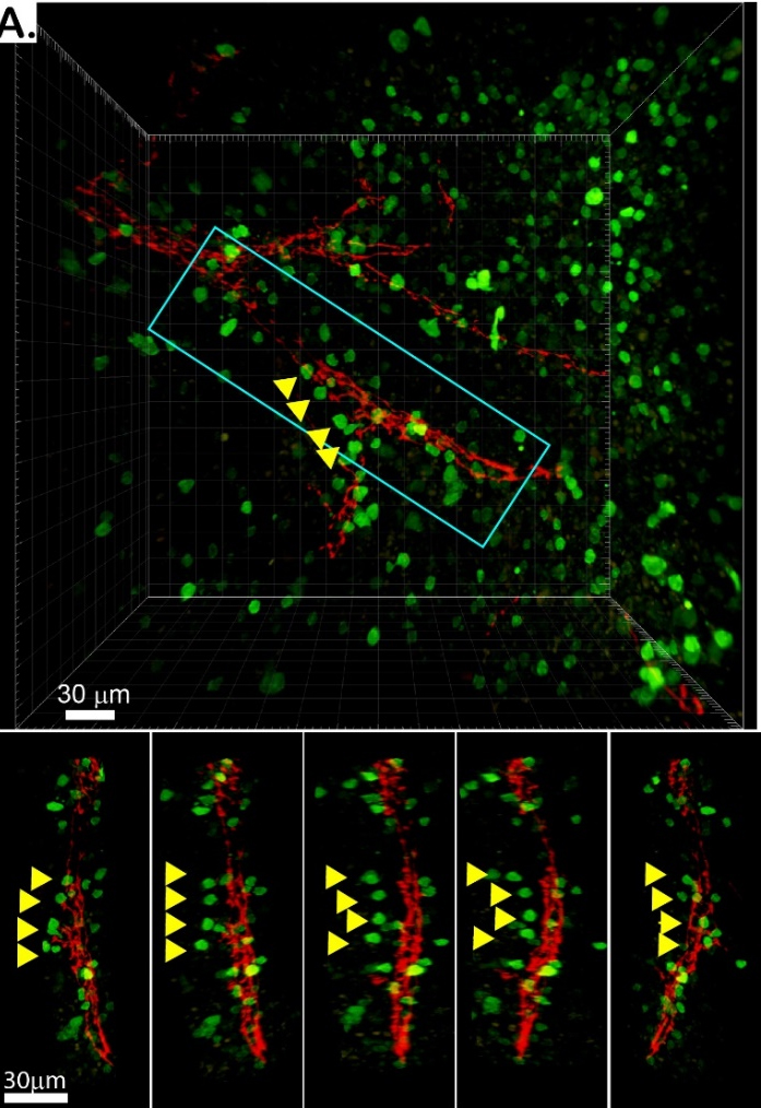
Innervation of lymphoid organs can profoundly alter immune cell function. The nervous system is an active participant during inflammation, reducing disease severity in conditions ranging from intestinal inflammation to septic shock.
Despite these seminal advances in neuro-immunology, there are significant gaps in our knowledge that impede the development of safe and effective therapeutics. High-resolution mapping of the relationships between neuron and immune cells has not been performed. This has led to debate over the anatomical origin of neurons and how to target immune reactions inn specific tissues. Resolving the origin of the neural signal will be important to activate this neuro-immune reflex for therapeutic applications.
Investigating pathogenic mechanisms of chemical-induced seizures
Colin Reardon
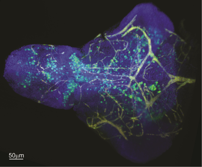
To investigate inflammatory mechanisms of chemical-induced seizures, double transgenic zebrafish larvae expressing farnesylated mCherry (mCherryF)-labeled macrophages and tumor necrosis factor alpha (tnfα) labeled with farnesylated enhanced GFP (eGFP-F) will be used to study macrophage activation in live animals. These data will require the ability to identify cell populations in three dimensions over time. It is hoped that these studies in combination with gene knockouts will identify chemicals and the receptors responsible for seizures.